心律失常患者跟踪计划

什么是心律失常?

What Is Arrhythmia?

An arrhythmia is an abnormal heart rhythm.It may feel like fluttering or a brief pause.It may be so brief that it doesn’t change your overall heart rate. Or it can cause the heart rate to be too slow or too fast. Some arrhythmias don’t cause any symptoms. Others can make you feel lightheaded or dizzy

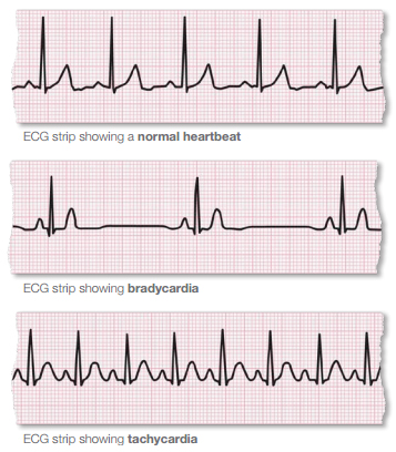
There are two basic kinds of arrhythmias.Bradycardia is when the heart rate is too slow — less than 60 beats per minute.Tachycardia is when the heart rate is too fast — more than 100 beats per minute.


What are the signs of arrhythmia?

  • When it’s very brief, an arrhythmia can have almost no symptoms. It can feel like a skipped heartbeat that you barely notice.
  • It also may feel like a fluttering in the chest or neck.
  • When arrhythmias are severe or last long enough to affect how well the heart works, the heart may not be able to pump enough blood to the body. This can cause you to feel tired, lightheaded or may make you pass out. It can also cause death.
  • Tachycardia can reduce the heart’s ability to pump, causing shortness of breath, chest pain,lightheadedness or loss of consciousness. If severe, it can also cause heart attack or death.

What are the signs of arrhythmia?

Before treatment, it’s important for your doctor to know where an arrhythmia starts in the heart and whether it’s abnormal. An electrocardiogram (ECG or EKG) is often used to diagnose arrhythmias. It creates a graphic record of the heart’s electrical impulses.Using a Holter monitor, exercise stress tests, tilt table test and electrophysiologic studies (“mapping” the electrical system of your heart) are other ways to find where arrhythmias start. Treatment may include:

  • Lifestyle changes
  • Medicine to prevent and control arrhythmias
  • Medicine to treat related conditions such as high blood pressure, coronary artery disease and heart failure
  • Anticoagulants to reduce the risk of blood clots and stroke.
  • A pacemaker to help your heart beat more regularly
  • Cardiac defibrillation and implanted cardioverter defibrillators (ICDs)
  • Cardiac ablation
  • Surgery

Non Shockable Rhythm’s

Non Shockable Rhythm’s

arrhythmias typesManagementEKG Strips

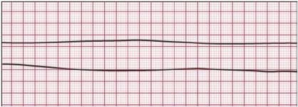
Asystole

  • Asystole is defined as a complete absence of electrical and mechanical cardiac activity.

Check two or more ECG leads for trace and amplitude

Commence CPR if no signs of life

Cannulate

Intubate

Adrenaline 1mg immediately then every 2nd cycle

Follow ARC guideline for non shockable rhythms (See Appendix)

Consider and correct 4 H’s and 4 T’s (see Appendix)

PEA is defined as any one of a heterogeneous group of organized electrocardiographic rhythms without sufficient mechanical contraction of the heart to produce a palpable pulse or measurable blood pressure.

Commence CPR if no signs of life

Cannulate

Cannulate

Intubate

Adrenaline 1mg immediately then every 2nd cycle

Follow ARC guideline for non shockable rhythms (See Appendix)

Consider and correct 4 H’s and 4 T’s (see Appendix)

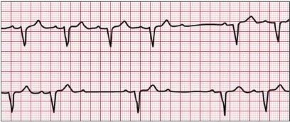
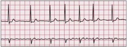
Bradycardias

Bradycardia is defined conservatively as a heart rate below 60 beats per minute, but symptomatic bradycardia generally entails rates below 50 beats per minute

Includes sinus bradycardia, heart blocks,idioventricular, and junctional rhythms

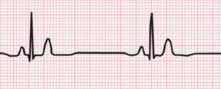
Heart rate of less than 60bpm

Normal p wave, QRS complex

Only if haemodynamically unstable Signs and symptoms of inadequate perfusion include hypotension, altered mental status, signs of shock, ongoing ischaemic chest pain, and evidence of acute pulmonary oedema

Atropine 500mcg up to 3mg

Consider 4H’s and 4 T’s

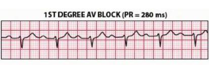
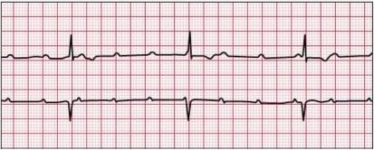
1st degree heart block

All p waves are conducted

PR interval greater than 0.20sec

There is no block just a delay in conduction

Only if haemodynamically unstable

Atropine 500mcg up to 3mg

Consider 4H’s and 4 T’s

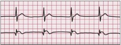
2nd degree Heart Block
Type 1 or Mobitz Ι or Wenckebach

Progressive delay of conduction of the AV node until conduction is completely blocked

PR interval is longer with each beat until QRS is dropped


Type 2, Mobitz ΙΙ

– 4 p waves before each QRS

Potential to progress to 3rd degree heart block

Ventricular rate less than atrial rate

If haemodynamically unstable;

Atropine 500mcg up to 3mg

Adrenaline 100mcg

Consider 4H’s and 4 T’s

Transcutaneous pacing

3rd degree Heart Block

No P waves are conducted

Disassociation between p wave and QRS complex

If haemodynamically unstable

Atropine 500mcg up to 3mg Adrenaline 100mcg

Consider 4H’s and 4 T’s

Transcutaneous pacing (See Appendix)

Junctional rhythm:

p wave often absent. "buried" in the QRS complex

p waves may be upside down or after the QRS

AV node has intrinsic automaticity that allows it to initiate and depolarize the myocardium during periods of significant sinus bradycardia or complete heart block

Treat underlying cause

Treat symptoms as for bradycardias (See Appendix)

Shockable Rhythm’s

Shockable Rhythm’s

arrhythmias typesManagementEKG Strips

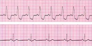
Unconscious/ Pulseless Ventricular Tachycardia (VT)

No detectable cardiac output Wide, regular QRS complex

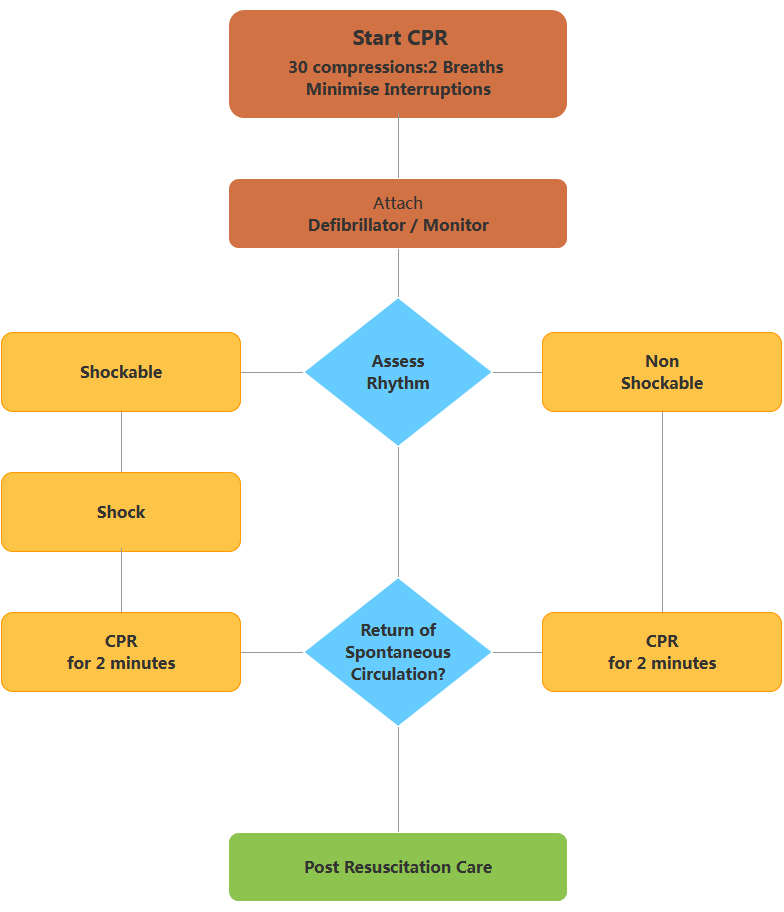
Commence CPR

Shock

CPR 2mins

Follow ARC guideline for Shockable rhythms (See Appendix)

Consider and correct 4H,s and 4T,s

Ventricular Fibrillation (VF)

no detectable cardiac output asynchronous ventricular activity rapid rate and disorganised with no uniform ventricular activit

Commence CPR

Shock

CPR 2mins

Follow ARC guideline for Shockable rhythms (See Appendix) Consider and correct 4H,s and 4T,s

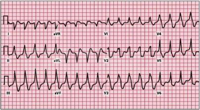
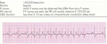
Supraventricular Tachycardia’s (SVT)

Tachycardia arising from atria or AV junction

Used to describe fast narrow-complex tachycardias

Usually caused by a re-entry circuit returning to the atria

ABC

Cannulate

Monitor haemodynamics

12 lead ECG

Treat reversible causes (see Appendix)

If haemodynamically unstable consider cardioversion

Refer to tachycardia algorithm (See Appendix)

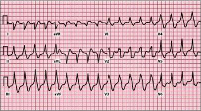
Rapid Atrial Fibrillation

Rate 100- upwards

Irregular rhythm

p waves fine or unable to see

Haemodynamically unstable

ABC

Cannulate

Monitor haemodynamics

12 lead ECG

Treat reversible causes (see Appendix)

If haemodynamically unstable consider cardioversion

Refer to tachycardia algorithm (See Appendix)

Conscious Ventricular Tachycardia

Usually regular, rate greater than 100

Wide or broad QRS complexes greater

than 3 small squares Patient is conscious

Patient has cardiac output

ABC

Cannulate

Monitor haemodynamics

12 lead ECG

Treat reversible causes (see Appendix)

If haemodynamically unstable consider cardioversion

Refer to tachycardia algorithm (See Appendix)

管理可逆的原因:4 H's

Management of Reversible causes: 4 H's

4HsMANAGEMENT
Hypox · Check and maintain airway
· Insert Guedel, ETT, LMA, surgical airway if required
·
Hypovolaemia · Replace blood or fluid loss Replacement of blood with:Crystalloid/ Colloid;Blood Products
· Anaphylaxis Management of ABC Adrenaline(IMI, S/C, or MV) Hydrocortisone Correct hypovolaemia.
Hypo/Hyperkalaemia Hypokalaemia
· Potassium of less than 3. 5mmol/L
· Replace Potassium
· K 5 mmol as slow bolus IV in severe hypokalemia Hyperkalaemia
· I calcium, 10 mLs 10% CaCI2, up to 3 ampoules, each over 5 minutes
· hyperventilation:Co2+H2O命H2Co3H++HCo3-
· 50mls 50 glucose 10 units Actrapid over 10-15 minutes
· NaHCO3 to correct acidosis
· Nebulised salbutamol
Hypo/Hypothermia Hypothermia

· Active core re-warming
· Warmed humidified oxyger
· Warmed intravenous fluids
· Peritoneal lavage
· Extracorporeal warming
· Pleural lavage Hyperthermia
· Cooling Blankets
· Cooling packs or ice to head, axilla, chest, groin and legs
· Cooled IV fluids

管理可逆的原因:4Ts

Management of reversible causes:4Ts

4TsMANAGEMENT
Tamponade · Pericardiocentesis
· open sternotomy wound if post cardiac surgery
Tension Pneumothorax Thoracocentesis
· Chest tube insertion if there is time or a large bore needle through the 2nd intercostal space in the mid-clavicular line
Toxins/tablets
· Antidote
· Charcoal (within 1 hr of ingestion)
· Supportive measures ABCDEFG
Hypo/Hypothermia
· Thrombolysis, embolectomy or cardiopulmonary bypass to allow operative removal of the clot

先进的技术给患者提供帮助

Advanced Lift Support for Adults

During CPR

Airway adjuncts(LMA/ETT)

Oxygen

Waveform capnography

Ⅳ/| O access

Plan actions before interrupting compressions

(e.g. charge manual defibrillator)

Drugs

Shockable

  • Adrenaline 1 mg after 2 shock then every 2 cycle
  • Amiodarone 300 mg after 3 shock

Non shockable

  • Adrenaline 1 mg immediately ten every 2nd cycle

Consider and correct

hypoxia

hypokalaemia

Hyper /hypokalaemia/metabolic disorders

Hypothermia/hyperthermia

Tension pneumothorax

Tamponade

Toxins

Thrombosis(pulmonary / coronary)

Post resuscitation Care

Re-evaluate ABCDE

12 lead ECG

Treat precipitating causes

Re-evaluate oxygenation and ventilation

Temperature control(cool)

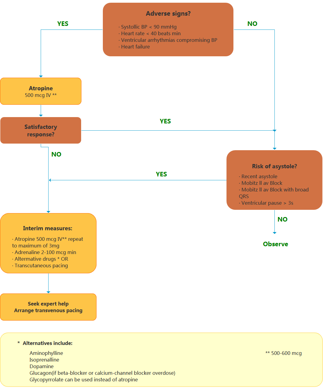
Bradycardia Algorithm

includes rates inappropriately slow for heamodynamic state

if appropriate,give oxygen,cannnulate a vein,and record a 12-lead ECG.

Tachycardia Algorlthm

(with pulse)